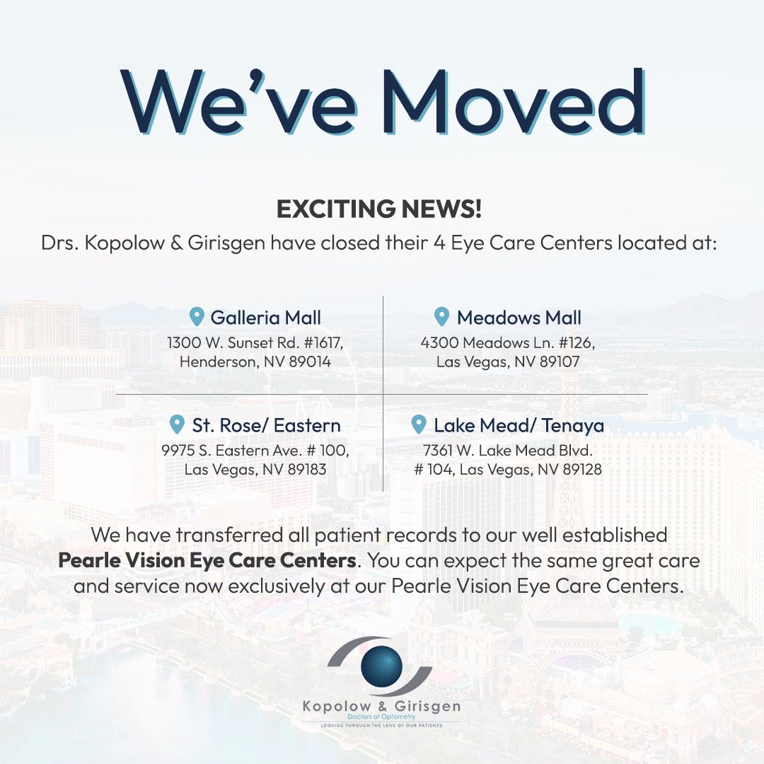
Helpful Articles

Helpful Articles

Helpful Articles

Every patient is different and so are their eyes. This means that there need to be different types of contact lenses to suit each individual. Some patients have corneal abnormalities which mean that conventional lenses won’t sit comfortably on the surface of their eyes, while others suffer from eye conditions that mean normal contact lenses won’t be comfortable or could irritate their eyes.
As you may have guessed from the name, specialty contact lenses are unconventional contacts that are designed for patients that regular contacts might not be suitable. Here are some of the main types of speciality contact lenses and who they are recommended for.
Who might be a good patient for specialty contact lenses?
Some of the patients that might benefit from specialty contact lenses include those who:
have been diagnosed with dry eye syndrome
have corneal scarring
have been diagnosed with keratoconus, a condition characterized by the bulging of the cornea
suffer from strabismus, a condition where the patient has an eye that turns in or out relative to the other
have suffered an injury to the eye
suffer from a peripheral corneal thinning disorder
are intolerant to other types of lenses
Your eye doctor or contact lens provider will be able to tell you if you need specialty contact lenses and if so, which lenses would be best based on your individual requirements.
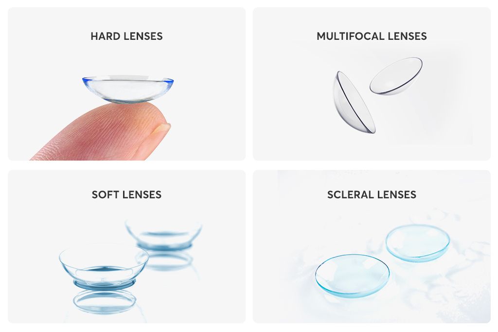
Rigid Gas-Permeable Lenses
Also known as RGP lenses, these are made from a special material that allows oxygen to pass through them and reach the surface of the eyes. This helps to keep the eyes hydrated and comfortable, making these lenses easier to wear, especially for patients who suffer from dry eyes. Dry eyes aren’t just a symptom, but a very real condition, characterized by dry, stiff, and uncomfortable eyes, blurred vision, and eye fatigue. RGP lenses are more rigid than soft lenses, and this helps to keep them stable and secure on the eyes so that patients can enjoy sharper vision. They also help the cornea to maintain its shape, which helps to minimize the effects of some corneal abnormalities.
Scleral Contact Lenses
Scleral contact lenses are very different to standard contact lenses. This is because scleral lenses are much larger in diameter, with three different sizes available depending on your specific needs. This size difference means that the edges of the contact lens fall on a white part of the eye, called the sclera rather than the cornea. Scleral lenses are also different in that they vault over the surface of the cornea rather than touching it, leaving a space between the front surface of the eye and the back of the contact lens. This makes scleral lenses a good choice for patients with dry eyes and corneal abnormalities. Space can trap tear film which keeps the eyes hydrated, while space also accommodates many corneal abnormalities, such as the bulge associated with keratoconus.
Limbal Fit Contact Lenses
Limbal contact lenses are another type of specialty lens that falls between rigid gas-permeable lenses and scleral varieties in terms of their size. Their larger overall diameter helps to increase their stability on the surface of your eyes. They also offer minimal interference with the eyelids, which helps to ensure comfort and clarity of your vision.
Hybrid Contact Lenses
Hybrid contact lenses are a combination of both soft and gas-permeable contact lenses, giving patients the opportunity to enjoy the best parts of both designs. The middle part of hybrid lenses is made from gas-permeable material that lets oxygen pass through to the eyes. However, the gas-permeable part of the lens is more rigid, and this firmer center gives the lens greater stability and the patient enhanced clarity. The RGP portion of the lens also helps to trap a tear film between the cornea and the lens so that the eye remains hydrated. Meanwhile, the outer edge of hybrid lenses is a soft lens skirt. This means that patients don’t have to deal with the hard edges associated with RGP lenses that may be uncomfortable. Instead, the comfort levels that patients experience are more like wearing fully soft lenses.
For more information about specialty contact lenses, don’t hesitate to speak to our dedicated eye care team.
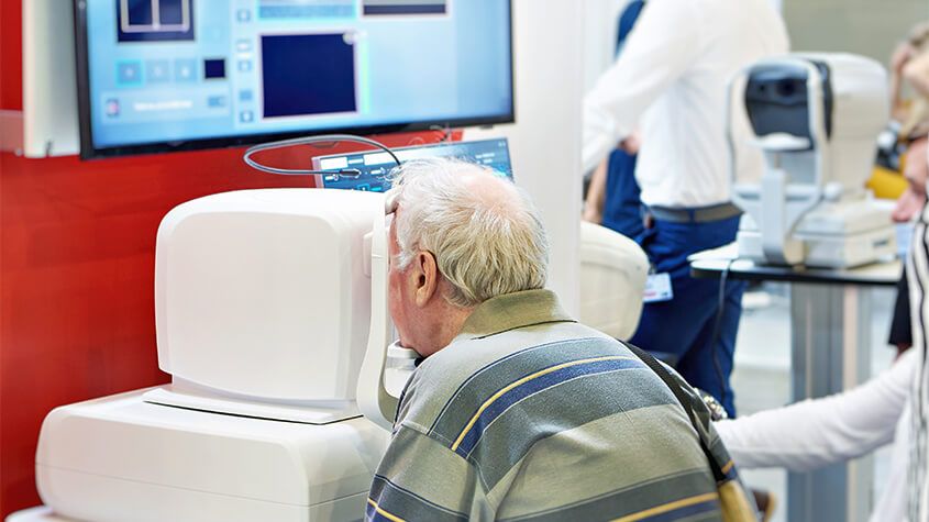
Optical Coherence Tomography is a non-invasive imaging test that may be performed as a standard part of your regular, comprehensive exams, or you may be able to request this test as an addition to your usual exam.
Optical Coherence Tomography uses light waves to take cross-section images of your retina, which is the area of light-sensitive cells at the back of your eye that is responsible for receiving light and transmitting it into messages that are sent up to the brain. The technology behind OCT enables your eye doctor to see each of the different layers that make up the retina. By being able to see these and measure them, they can obtain a much clearer picture of the overall health and condition of your eyes.
Why are Optical Coherence Tomography scans important?
When you choose to have an OCT scan at fairly regular intervals, such as during your normal comprehensive eye exams, your eye doctor can compare newer results to previous ones. This helps them to build up a picture of the health of your eyes, and spot any changes which may be concerning, early, before they cause symptoms or have a permanent effect on your vision.
Anyone can have an OCT scan, but they are particularly recommended for patients over the age of 25 who are concerned about the health of their eyes, or who are at risk of or already have diabetes, glaucoma or a family history of eye disease. This is because they can be used to spot the early signs of a range of eye diseases, including glaucoma, diabetic retinopathy, macular degeneration, disorders of the optic nerve and more – even before you realize that you are affected.
What happens during an Optical Coherence Tomography scan?
An OCT scan is a quick, painless experience. To prepare you, your eye doctor may require you to have eyedrops that will dilate your pupils and make it easier to see your retina. This means that the scanner will get clearer, more concise images. You’ll be asked to sit in front of the OCT machine where you will rest your head against a support to help you sit perfectly still. As you stare ahead, the equipment will perform the scan of your eyes. There is no contact with your eyes whatsoever, you will just need to sit still, with your eyes open as much as possible during the process, which usually takes less than 10 minutes. The images will be sent digitally to your eye doctor for them to assess immediately and stored digitally on your personal record.
There’s no downtime after an OCT scan, but if you have had your eyes dilated you may find that you are particularly sensitive to light for a few hours afterwards. This occurs because the pupils remain wider and therefore able to let more light in that usual.
If you would like to find out more about Optical Coherence Tomography, don’t hesitate to speak to our professional eyecare team.

If you find it difficult to tell colors apart, you may be color blind. Color blindness, or color deficiency, is estimated to affect around 8% of men and about 1% of women, but for those affected, it can significantly impact the quality of their day-to-day life. Contrary to popular belief, being color blind doesn’t mean that you can’t see any color at all. Instead, patients simply struggle to differentiate between certain colors. The vast majority of people who are color blind find it impossible to tell the difference between varying shades of red and green. You may hear this referred to as red-green color deficiency. However, this doesn’t only mean that they mix up red and green. They can also mix up colors that have some green or red light as part of their whole colors, for example purple and blue. This is because they are unable to see the red light that forms part of the color purple.
As you can probably imagine, this type of visual impairment can be a problem for things like traffic lights, taking medications and even looking at signs and directions. For example, someone who is color blind may find that the green on a traffic light may appear white or even blue.
EnChroma lens technology is specifically designed to counteract red-green color deficiency and enable patients to better identify the difference in these colors or shades. They do this by selectively filtering out the red and green wavelengths of light at the exact point where the color sensitivities overlap before hitting the retina, creating far greater contrast between the colors so that the patient can distinguish between them successfully. Most cases of color blindness respond well to EnChroma’s innovative spectral lens technology, giving patients the ability to experience life in bright, vibrant technicolor.
EnChroma lenses are made from leading edge, Trivex material, and this helps to give them the best possible quality and clarity of vision. These lenses are also extremely light, strong and offer patients 100% protection against UV light, helping to keep your eyes healthy as well as improving your vision.
If you or someone you know is color blind or color deficient and could benefit from EnChroma lenses, contact us today to learn more about how they can help!

Wearing contact lenses gives patients the flexibility and freedom to live life to the fullest, without some of the difficulties presented by wearing glasses. Many people who choose contact lenses do so because they don’t like the way that glasses look or feel, or because wearing glasses compromises their ability to perform certain tasks or activities, such as sports or jobs that require the use of safety goggles.
There are lots of different contact lenses to choose from, with two of the most popular being daily disposables and toric lenses.
Disposable Lenses
As their name suggests, these daily contact lenses are disposable. This means that they can and should be discarded at the end of each day rather than re-worn. Disposable lenses do tend to be a little more expensive than some repeat-wear varieties, but the benefits usually outweigh the cost.
Some of the advantages of choosing daily disposable contact lenses include:
You don’t have to clean them, which saves patients a great deal of time and hassle. It also helps save money in terms of the ongoing cost of cleaning solution.
Disposable lenses are also great for people with eye allergies. This is because with ordinary lenses, there’s an opportunity for deposits and microorganisms to build up. With daily disposables, allergens have less chance to attach themselves to the lenses and cause irritation and other allergy symptoms.
You don’t need to schedule regular replacements either, which makes wearing contact lenses easier on your schedule.
Disposable contact lenses are particularly good for people who have busy lives and are likely to cut corners when it comes to caring for their eyes or contacts since there is no cleaning or maintenance required.
Daily disposable contact lenses are available in a wide range of prescriptions, including those for patients with nearsightedness and farsightedness. Your eye doctor will be able to advise you if you are a candidate for disposable contact lenses.
Toric Lenses
Toric contact lenses are recommended for patients who have a refractive eye problem called astigmatism. Patients with astigmatism have corneal abnormalities that cause the refraction of the eye to be different between the vertical and horizontal planes, causing blurred vision and difficulty seeing fine details. Toric contact lenses are shaped in a particular way that creates the different focusing powers needed in each part of the lens to correct your vision. For this reason, it’s essential that Toric lenses are placed into the eyes in the correct position. Fortunately, manufacturers design Toric lenses with features that help them to stay in place, including:
Thin/thick zones
Creating areas of the lens that are thicker or heavier which helps secure it in position
An area where the bottom of the lens is slightly cut off
To keep them stable, Toric lenses are a little firmer than conventional soft lenses. This means that some patients can find them a little less comfortable, but the superior vision they obtain outweighs this. Your eye doctor will be able to advise you if you are a good candidate for Toric contact lenses and which variety would best suit you.
To find out more about daily contact lenses, speak to our friendly and knowledgeable team.
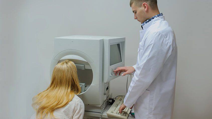
Visual field testing is an important part of most standard comprehensive eye exams. Also sometimes known as perimetry testing, Visual field testing is a method to measure the entire scope of vision of an individual, including their peripheral/side vision.
The importance of visual field testing
Visual field testing is one of the most effective diagnostic treatments in the detection of glaucoma. This is because when patients are affected by glaucoma, it is usually the peripheral vision that is affected by their condition first. However, it can also be used to detect central or peripheral retinal diseases, eyelid conditions such as drooping, optic nerve damage and conditions that affect the visual pathways from the optic nerve to the area of the brain where this information is processed into vision.
Visual field testing is also an important part of monitoring for people who are considered to be at risk for vision loss from disease and other problems, including those who have been diagnosed with the following:
- Multiple sclerosis
- Hyperthyroidism
- Pituitary gland disorders
- Central nervous system problems (such as a tumor that may be pressing on the brain)
- Stroke
- Diabetes
- High blood pressure
What to expect from visual field testing
There are a variety of methods that can be used to perform visual field testing, including:
Static automated perimetry. This is where a machine is used to quantify how well the patient is able to detect flashing lights of varying size and brightness in different areas of their visual field, while they concentrate on a central point. The patient responds by pushing a button when they see the light.
Kinetic perimetry. This involves points of light that are fixed in size and intensity and are presented along the patient’s peripheral vision, before being gradually moved inwards to determine their field of vision.
Visual field testing is non-invasive, painless and doesn’t require patients to have their eyes dilated. The results, which are usually presented in a series of charts, are digital and sent directly to your eye doctor for interpretation. Depending on the outcome of your results, you may be recommended for further diagnostic testing which could include blood tests. If you have been diagnosed with glaucoma, you will probably be recommended to have several visual field tests each year, which will help your eye doctor to monitor the progression of your condition and recommend treatments to slow it.
If you would like more information about visual field testing, or if you have concerns about your peripheral vision, please don’t hesitate to schedule an appointment with our experienced and knowledgeable eyecare team today.

Optomap is an innovative new technology that gives eye doctors the ability to perform ultra-wide retinal imaging that is far superior to what can currently be achieved using conventional retinal imaging options. In contrast to conventional retinal imaging, Optomap captures at least 50% more of the retina in a single capture, and with Optomap’s multi-capture function, up to 97% of the retina can be viewed. This gives eye care professionals greater opportunity to monitor the health and condition of patient vision.
Why is Optomap important?
Optomap is another great preventative eyecare technology tool. By allowing your eye doctor to have a comprehensive view of your retina, they will be able to detect any developing eye diseases early on, before they have a detrimental impact on your vision and day to day life. Not only can Optomap detect eye conditions such as retinal holes, retinal detachment, macular degeneration and diabetic retinopathy, but it can also be used to identify some general health conditions such as cardiovascular disease, stroke and cancer.
What to expect from Optomap scanning
Optomap is a fast, painless and non-invasive procedure that is suitable for patients of all ages, even children and pregnant women. Many patients require their eyes to be dilated ahead of the scan and will be given eyedrops which will widen their pupils and make it easier for the camera to see the structures inside the eye. Pupil dilation is painless, but patients may feel more sensitive to light both during their Optomap scan and afterwards for up to 24 hours. You may also have slightly blurred vision for a few hours. Once your eyes are dilated, you’ll be sat down and asked to look into a small device that will take the pictures of your retina. A short flash of light will let you know that the image has been taken, and the entire imaging is over in just a few seconds. The results will be sent digitally to your eye doctor who will then evaluate them. The results will also be stored on your personal optical record for future information.
If you would like more information about what is involved in Optomap, or to schedule an appointment for this effective screening technology, please contact our eyecare team.
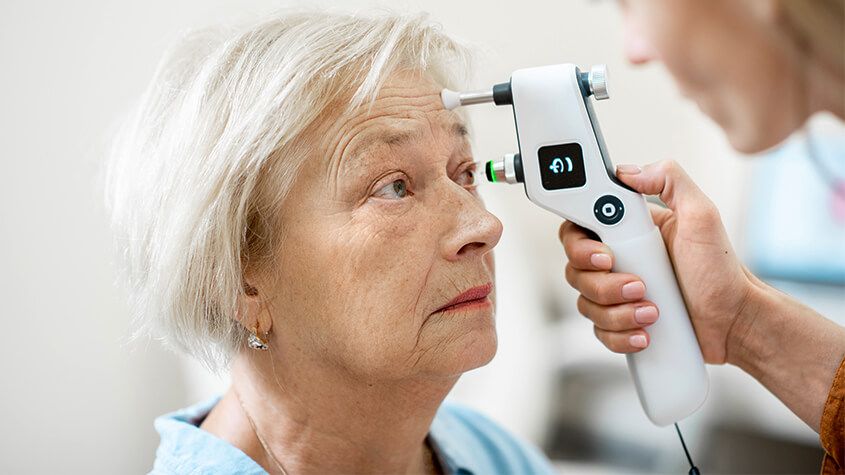
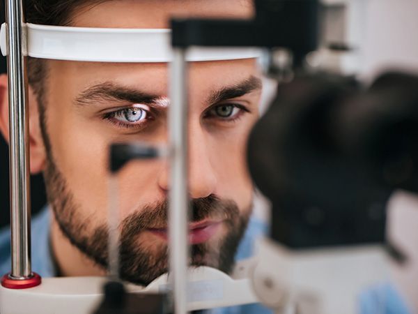
A tonometer refers to the equipment that is used in tonometry – a test that measures the pressure inside your eyes, also known as intraocular pressure or IOP for short. Tonometry is rarely performed at your average comprehensive eye exam unless you are at high risk of or have been already diagnosed with glaucoma. Fortunately, tonometry can be used to detect changes in eye pressure before they cause any symptoms, enabling prompt action to be taken before your vision is affected.
About glaucoma
Glaucoma is a common eye condition that occurs when the optic nerve, which connects the eye to the brain, becomes damaged. It’s normally caused by fluid building up in the front part of the eye, which causes the pressure inside the eyes to build. As the pressure increases, the optic nerve becomes increasingly damaged, and this prevents messages from being transmitted between your eyes and brain effectively. As a result, the patient’s vision becomes compromised. Without treatment, the level of vision loss will continue to increase. Unfortunately, any vision that has been lost as a result of glaucoma cannot be restored.
Most of the time, glaucoma develops very slowly which means that many people don’t realize that they are affected until some damage to their vision has already occurred. However, occasionally glaucoma can develop quickly, and symptoms do occur. These can include:
- Red eyes
- Intense headaches
- Tenderness around the eyes
- Eye pain
- Seeing rings/halos around lights
- Blurred vision
- Nausea and vomiting
If you notice any of these symptoms, it’s important that you make an appointment with your eye doctor right away so that you can be assessed. You are likely to have a tonometry test as part of this assessment.
What to expect from tonometry testing
There are various methods of tonometry testing, but many eye doctors use either Goldmann tonometry, which is the conventional technique to measure eye pressure, or electronic tonometry.
Goldmann tonometry testing is carried out using the Goldmann applanation tonometer, which is attached to a slit lamp microscope. This requires anesthetic eye drops to be used which numb your eyes, before a small probe is pressed gently against the eye, indenting the cornea. The pressure that the cornea pushes back onto the tonometer is what is measured to give your IOP reading. Electronic tonometry is where a handheld, mobile device is gently and quickly applied to the cornea to check the pressure, providing an accurate reading. Some eye doctors also offer non-contact tonometry which is where a puff of air is used to flatten the cornea, although this is reported to be less accurate than the Goldmann technique.
If you would like to find out more about Tonometry testing, please call our office to speak with our dedicated eyecare professionals.
We all want to look our best and in the last decade, we have seen a significant increase in the number of people seeking cosmetic services in order to enhance their appearance. With our eyes being our most distinguishing feature, we want to make the most of them. Thankfully there is now a range of cosmetic services that can help to rejuvenate our eyes and the area around them to keep them fresh, young and wrinkle-free.
Let’s take a look at some of the services on offer.
Pigment removal
The brown pigment spots that appear on the face are often referred to as age spots and are a result of sun exposure. With age, the repeated exposure to UV rays causes melanin, a compound that is responsible for pigmentation and protecting the skin begins to clump together to form an area of hyperpigmentation. Whilst they aren’t any cause for concern, many people feel that they look unsightly. Luckily, there are a number of different treatments that you can get to remove them including topical creams, laser therapy, and chemical peels.
If you are suffering from darker pigmentation then we strongly recommend that you make an appointment with a qualified dermatologist who will recommend the best course of treatment for you, based on your specific needs.
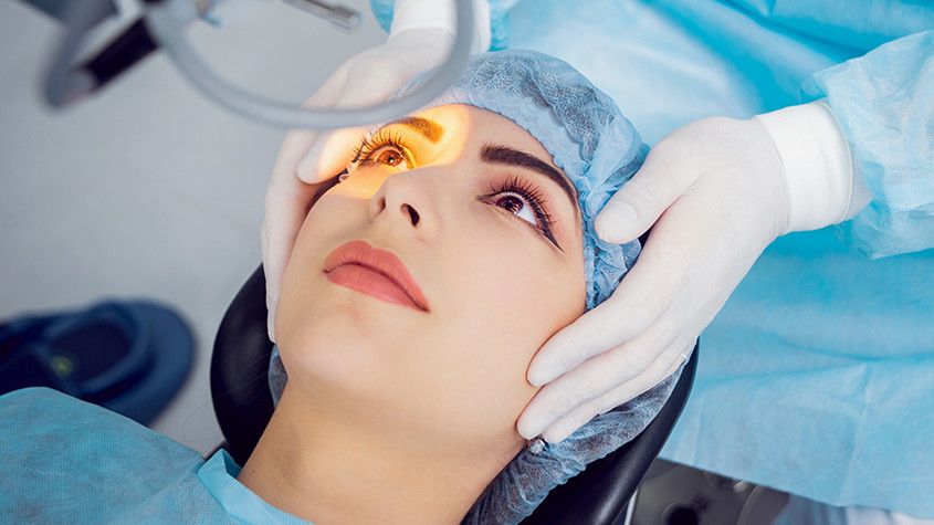
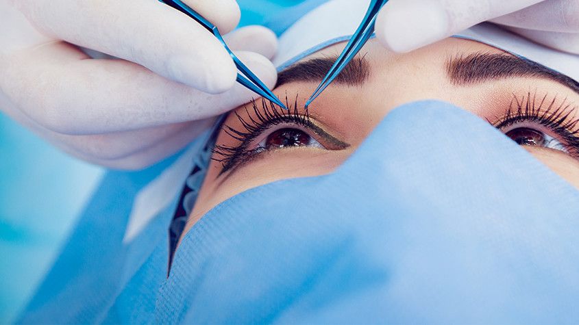
If you are one of the thousands of people considering LASIK laser eye surgery, then you will probably be gathering as much information as possible about the treatment. By this point, you are probably aware of the benefits that LASIK offers, such as a reduced or eliminated need for glasses or contact lenses and greater convenience in your day to day life. However, for many patients, despite the advantages of LASIK, the thought of surgery on their eyes is still a cause of anxiety and fear. One of the best ways to alleviate this concern is to find out more about what the procedure entails.
Your consultation
Before you can be approved for any form of laser vision correction, including LASIK, you will need to attend a consultation appointment with your surgeon. During the consultation, he will perform an examination of your eyes and use your medical and ocular history to determine if you are a good candidate for the procedure. He will also speak to you about the expected outcome from your surgery, making you aware that while LASIK will dramatically improve your eyesight, there is no guarantee that you will not need to wear glasses in some situations, such as while driving in the dark.
How LASIK Works
LASIK uses a cool, ultraviolet beam of light to reshape the patient’s cornea. Doing so will more accurately focus the light that enters the eye on to the retina, thus improving the patient’s vision. The way in which the cornea needs to be reshaped will depend on the visual needs of the patient. For example, a patient who is far-sighted will need their cornea reshaping to be steeper to experience better eyesight. Alternatively, a patient who is near-sighted will require their cornea to be flattened in order to improve their vision. LASIK can also smooth an irregular cornea into a more standard shape, meaning that the procedure can also be used to correct astigmatism.
The LASIK procedure
The LASIK procedure is very fast and straightforward. Although you will probably be in the surgical suite for around half an hour, the actual process only takes a couple of minutes per eye. The rest of the time will be spent preparing and ensuring that you are comfortable. Anesthetic eye drops are given to patients before their procedure so that the entire process is pain-free. If you are particularly anxious, it may also be possible for you to be slightly sedated – this should be discussed with your doctor at your consultation appointment.
Once you are in position, we will use a femtosecond laser to cut a thin, circular flap into the outer cornea. This can then be pulled back to reveal the underlying corneal tissue, known as the stroma so that it can be reshaped using the laser. The exact path that the laser needs to take, known as the topography, will have been pre-programmed ahead of the procedure and can be followed with complete precision and accuracy.
Once the reshaping is complete, the flap is replaced back over the eye and the surgery is complete. There is no need for sutures or bandages as the cornea will start to heal immediately and without any medical intervention.

If you already rely on wearing glasses or contact lenses to be able to see clearly, you may be frustrated with the effect that they have on your life. Regular vision tests, finding glasses to suit your face shape, having to remember to take eyeglasses with you wherever you go, prescription sunglasses, fiddly contact lenses… the list of inconveniences associated with conventional ocular solutions is extensive.
LASIK is a modern, minimally-invasive procedure that can substantially reduce or eliminate your need to use eyeglasses or contact lenses, allowing you to enjoy life without limitations or inconvenience. The popularity and success of LASIK laser eye surgery have helped to make it the number one elective surgery across the globe.
Candidacy for LASIK
LASIK has an extremely high success rate. According to the American Society of Cataract and Refractive Surgery, 96% of patients achieve 20/20 vision or better. However, it’s high success rate doesn’t make LASIK automatically the right solution for everyone.
Candidacy for LASIK is assessed by our doctors on a case by case basis so that you be certain that whatever treatment is recommended for you, it will give you the very best opportunity to improve your vision. During your consultation, our doctors will perform a thorough examination of your eyes and vision, ask you about your general health and talk you through both the procedure and aftercare.
The general guidelines for LASIK candidacy state that patients must:
be at least 18 years of age
have had stable vision with no prescription changes for a minimum of 12 months
have a current prescription for eyeglasses or contact lenses that falls between specified parameters (Our doctors will be aware of what these parameters are)
have no significant medical or eye-related problems such as glaucoma, macular degeneration or diabetic retinopathy
have no history of corneal disease
not be pregnant or nursing at the time of the procedure

Powered by: